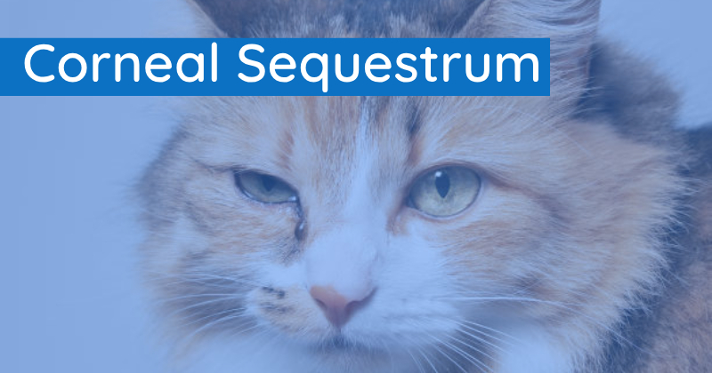
Corneal Sequestrum Conundrum

What is it?
A corneal sequestrum is a layer of dead tissue on the surface of the cornea (outer layer of the eye) that is typically dark brown or black. This is a painful condition that has the potential to become infected, leading to eye rupture and possible loss of the eye.
What causes it?
Sequestrums are often formed following a corneal ulcer. While the exact cause as to why they form is unknown, it appears to be breed-related and/or due to infectious factors.
How is it treated?
The recommended treatment is surgery to remove the dead layer of tissue and repair the defect with a graft. This is a day surgery performed at high magnification with the aid of an operating microscope. The third eyelid is then held across the eye for two weeks with a stitch to help protect and aid in the healing of the cornea.
What to expect after surgery?
Patients will be discharged with eye drops as well as oral medications and an e-collar will be placed. A post-operative check is scheduled for two weeks after the surgery, at which time the third eyelid suture is removed, and the eye is examined. Further medications will then be prescribed as needed. The success rate of the surgery is around 95%, though occasionally, the sequestrum can recur.
How can we help?
Our experienced veterinary team can diagnose and perform high-level corneal sequestrum repair surgery.
Want to know more about our ophthalmology services?
